
Abstract 13
Response rate of ipsilateral corticospinal TMS
Sousa, Maria Helena Gomes 1; Fonseca Junior, Paulo Roberto 1; Marchiore, Patrícia Monteiro 1; Battistella, Linamara Rizzo 1; Fregni Felipe 2 ; Simis, Marcel 1
- Physical and Rehabilitation Medicine Institute, General Hospital, Medical School of the University of Sao Paulo, Sao Paulo, Brazil; 2. Neuromodulation Center and Center for Clinical Research Learning, Spaulding Rehabilitation Hospital and Massachusetts General Hospital, Harvard Medical School, Boston, Massachusetts, USA
OBJECTIVE: To describe and compare the contralateral and ipsilateral neurophysiological evaluation with TMS of healthy participants
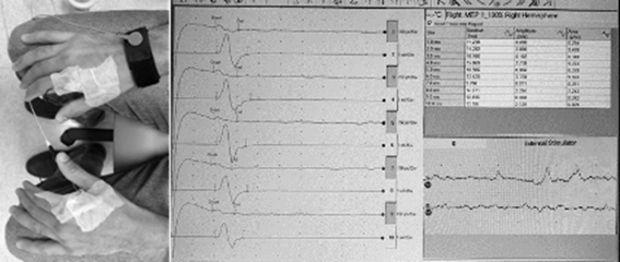
METHODS: 100 healthy participants were included (CAAE 76850117.3.0000.0068). Neurophysiological TMS was performed with measurement of the Motor Evoked Potentials (MEP) of both hemispheres. The response to the stimulus was monitored by electromyography on muscles of both hands (first dorsal interosseous) in order to assess the contralateral and ipsilateral corticospinal tract.
RESULTS: There was an ipsilateral motor response in 4 of the 100 examined individuals. We found a low MEP amplitude of the ipsilateral pathway (mean of 0.12 mV) when compared to the contralateral pathway of the same participant (mean 1.97 mV), however with similar shape and latency of the wave.
CONCLUSIONS: This study found a 4% response in the ipsilateral tract. There was a reduced amplitude of the ipsilateral response, this is probably related to the smaller number of bundle of fibers compared to the contralateral side. This study suggests that the presence of ipsilateral pathways is rare, however, it should be investigated better since it can influence rehabilitation of brain injuries.
FIGURE
ACKNOWLEDGMENTS: I would like to thank everyone involved, such as Karin, Artur, Karinna, Natalia,Christiane and Simon.
FUNDING/FINANCIAL SUPPORT: This work is supported by a FAPESP grant (SPEC 2017/12943-8)
DOI: https://doi.org/10.21801/ppcrj.2020.S1.13