
Abstract 25
Astrocitary Response In Vitro to Static Magnetic Stimulation
Ronaldo F Castanho¹; Sara F Nunes¹; Ricardo N Goulart¹; Caroline C da Costa¹; Izabel C de Souza¹.
- Cell Modulatory Laboratory – Institute of Biology - Department of Morphology - Federal University of Pelotas - Pelotas, Rio Grande do Sul, Brazil.
OBJECTIVE: Search for the possible morphological and biochemical effects of static magnetic stimulation in primary astrocyte culture.
METHODS: The astrocytes of the cerebral cortex of neonate Wistar rats were seeded in 24 well plates with growth medium, changed every 3 or 4 days. After 18 days of incubation at 37ºC and 5% CO2, the cells were divided into 4 groups: control (C), which was not stimulated, and groups (5), (15) and (30) stimulated by 5, 15 and 30 min respectively, for 7 days. The concentration of reactive oxygen species (ROS) was quantified; the cells morphology was analyzed by photographic recording in the optical microscope; while the protein content was verified by the sulforodamine B assay.
RESULTS: There were no morphological changes in any group (Figure 1.A). Groups (5) and (15) had their cell density increased (Figure 1.C), which may be associated with hypertrophy and hyperplasia of astrocytes - a replying mechanism of cellular damage. Finally, in group (5) there was a significant increase in the production of ROS (Figure 1.B), when compared to the control group. This finding could be explained by the cellular adaptation to the stimulus tending to homeostasis.
CONCLUSIONS: After the treatment period, there was no difference in cell morphology, but cell density and ROS concentration showed changes. Therefore, it is necessary to evaluate other parameters of static magnetic stimulation to understand the mechanisms of this stimulation on the functionality of astrocytes.
FIGURE. Effect of static magnetic stimulation on primary astrocyte culture.
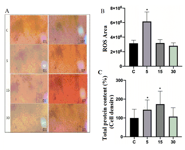
- A) Images obtained under an Inverted Light Microscope captured on the 1st (D1) and on the 7th day of static magnetic stimulation (D7); 400X magnification. B) Graph of cell density by total protein content in astrocytes. * Significantly different from the control: 5 min (p = 0.025) and 15 min (p <0.0001). C) Graph of production of reactive oxygen species in astrocytes. * Significantly different from the control (p <0.0001). Groups: C (control), 5 (5 min of stimulation) 15 (15 min of stimulation), 30 (30 min of stimulation).
KEYWORDS: static magnetic stimulation, astrocytes, morphology, biochemistry.
ACKNOWLEDGMENTS: Federal University of Pelotas (UFPEL), Laboratory of Immunology and Veterinary Virology (LABVIR), Laboratory of Cell Neuromodulation (NEUROCELL), Bioengineering of Hospital de Clínicas de Porto Alegre (HCPA).
FUNDING/FINANCIAL SUPPORT: Universal Call of the National Council for Scientific and Technological Development (CNPQ), Research Support Institute of Rio Grande do Sul - FAPERGS, and Funder of Studies and Projects (FINEP)
DOI: https://doi.org/10.21801/ppcrj.2020.S1.25